What is an MRI?
Magnetic resonance imaging (MRI) is a non-invasive medical test that utilises a powerful magnetic field, radio waves and a computer to produce detailed pictures of the internal structures of the body. Unlike conventional x-ray examinations and computed tomography (CT) scans, MRI does not utilise ionizing radiation. Instead, radio waves are used to temporarily change the alignment of hydrogen atoms that exist naturally within the body. As the hydrogen atoms return to their usual alignment, they emit energy that varies according to the type of body tissue from which they come. The scanner captures this energy and creates a picture of the tissues scanned based on this information.
Frequently, the differentiation of abnormal (diseased) tissue from normal tissues is better with MRI than with other imaging modalities such as x-ray, CT, and ultrasound, especially for the brain, spine, muscles and joints.
Enhance Patient’s MR Imaging Experience

Optimized Resolution

Reduced Anxiety

Soothing Music
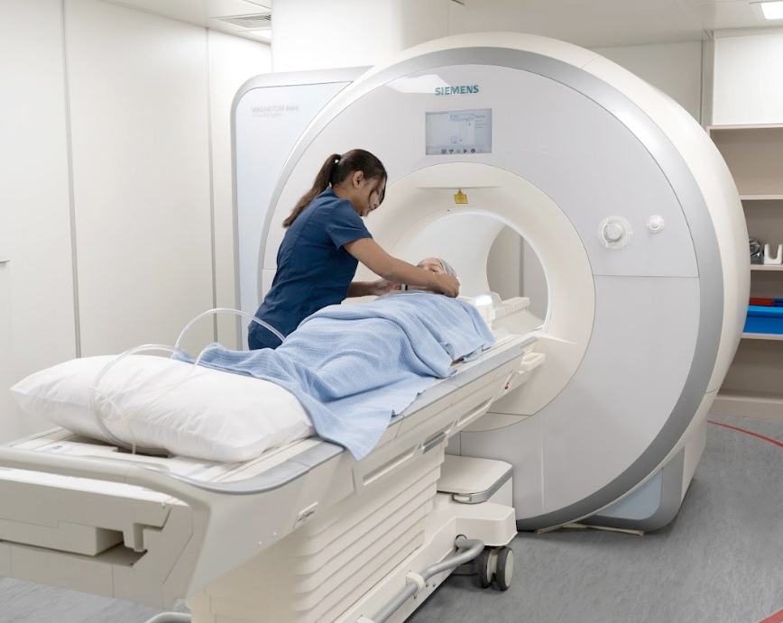
- Siemens’ 70cm wide and short bore MR system provides better
- patient comfort with its large size and wide opening bore
- Reduce potential claustrophobia and anxiety
- Available at all Lifescan Imaging Centres

How is an MRI Performed?
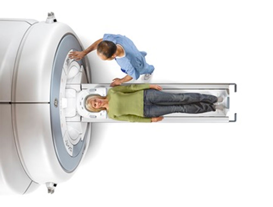
You will lie down on top of a moveable examination table. Padded straps may be used to help you stay still and maintain the correct position during imaging. Devices that contain equipment for sending and receiving radio waves may be placed around or next to the area of the body being studied. You will be placed onto the magnet of the MRI unit and the radiologist will perform the examination while working from a computer outside of the room.
If a contrast needs to be used for the scan, an intravenous catheter, also known as an IV line, will be inserted into a vein in your hand or arm. The intravenous needle may cause some discomfort when it is inserted and you may experience some bruising. Some patients may experience a temporary metallic taste in their mouth after the contrast injection. Your intravenous line will be removed after the scan.
MRI exams generally include multiple runs (sequences), some of which may last several minutes. Depending on the type of examination, the whole procedure is usually completed in 30 to 50 minutes. When the examination is complete, you may be asked to wait until the technologist checks the images in case additional images are needed.

What will I Experience During and After an MRI?
Most MRI exams are painless. However, some patients find it uncomfortable to remain still during MR imaging. Others experience a sense of being “closed-in” (claustrophobia). Sedation can be arranged for patients who anticipate anxiety, but fewer than one in 20 require medication.
It is normal for the area of your body being imaged to feel slightly warm, but if it bothers you, notify the radiologist. It is important that you remain perfectly still while the images are being obtained, which is typically only a few seconds to a few minutes at a time. You will know when images are being recorded because you will hear and feel loud tapping or thumping sounds when the coils that generate the radiofrequency pulses are activated. Earplugs or headphones (and music) to reduce the intensity of the sounds made by the MRI machine can be provided.
You will usually be alone in the exam room during the MRI procedure. However, the technologist will be able to see, hear and speak with you at all times using a two-way intercom. A friend or parent may stay in the room as long as they are also screened for safety in the magnetic environment.
If you have not been sedated, no recovery period is necessary. You may resume your usual activities and normal diet immediately after the exam. On very rare occasions, a few patients experience side effects from the contrast material, including nausea and local pain. Similarly, patients are very rarely allergic to the contrast material, but if they do, they can possibly experience hives, itchy eyes or other reactions. If you experience allergic symptoms, notify the technologist. A radiologist or doctor will be available for immediate assistance.
How should I Prepare for an MRI?
Clothing and Accessories
- You may be asked to wear a gown during the exam or you may be allowed to wear your own clothing if it is loose-fitting and has no metal fasteners.
- Jewelry and other accessories should be left at home if possible, or removed prior to the MRI scan, as they can interfere with the magnetic field of the MRI unit, metal and electronic items are not allowed in the exam room. These items include:
- Jewelry, watches, credit cards and hearing aids, all of which can be damaged
- Pins, Hairpins, Metal Zippers and similar metallic items, which can distort MRI images
- Removable Dental Work
- Pens, Pocket Knives, and Eyeglasses
- Body Piercings
Diet Preparation
Guidelines about eating and drinking before an MRI exam vary with the specific exam being performed. Unless you are told otherwise, you may follow your regular daily routine and take food and medications as usual.
Declaration of Allergies and Health Problems
- Some MRI examinations may require you to receive an injection of contrast material into the bloodstream. The technologist or a nurse may ask if you have allergies of any kind, such as an allergy to iodine or x-ray contrast material, drugs, food, or the environment, or if you have asthma. The contrast material most commonly used for an MRI exam contains a metal called gadolinium. It is far less common for a patient to have an allergy to a gadolinium-based contrast agent used for MRI than the iodine-containing contrast for CT. However, even with known allergies to gadolinium, it may still be possible to use it after appropriate pre-medication. Patient consent will be requested in this instance.
- You should also let us know if you have any serious health problems, or if you have had any previous surgery. Some conditions, such as severe kidney disease, may prevent you from being given gadolinium contrast for an MRI and alternative arrangements can be made.
- If you have claustrophobia (fear of enclosed spaces) or anxiety, you may want to ask your doctor for a prescription for a mild sedative prior to your scheduled examination.
Pregnant Women and Children
Pregnant Women
If you are female, you should always inform your doctor or radiologist if there is any possibility that they are pregnant. MRI has been used for scanning patients since the 1980s with no reports of any ill effects on pregnant women or their unborn babies. However, because the unborn baby will be in a strong magnetic field, in general pregnant women should not have this exam in the first trimester of pregnancy unless the potential benefit from the MRI exam is assumed to outweigh the potential risks. Pregnant women should also not receive injections of gadolinium contrast material except when absolutely necessary for medical treatment.
Children and Infants
Infants and young children usually require sedation or anesthesia to complete an MRI exam without moving. Whether a child requires sedation will depend on the child’s age and the type of exam being performed. Special arrangements can be made in these cases.
Medical Implants, Devices and Foreign Bodies
Implants
In most cases, an MRI exam is safe for patients with metal implants, except for a few types. People with the following implants cannot be scanned and should not enter the MRI scanning area:
- Cochlear (Ear) Implant
- Some Types of Clips Used for Brain Aneurysms
- Some Types of Metal Coils Placed Within Blood Vessels
- Nearly All Cardiac Defibrillators and Pacemakers
In general, metal objects used in orthopedic surgery pose no risk during MRI. However, a recently placed artificial joint may require the use of another imaging procedure. If there is any question of their presence, an x-ray may be taken to detect and identify any metal objects.
Medical Devices
You should tell the technologist if you have any medical or electronic devices in your body. These objects may interfere with the exam or potentially pose a risk, depending on their nature and the strength of the MRI magnet. Many implanted devices will have a pamphlet explaining the MRI risks for that particular device. If you have the pamphlet, it is useful to bring that to the attention of the radiologist or scheduler before the exam. Some implanted devices require a short period of time after placement (usually six weeks) before being safe for MRI examinations. Examples include but are not limited to:
- Artificial Heart Valves
- Implanted Drug Infusion Ports
- Artificial Limbs or Metallic Joint Prostheses
- Implanted Nerve Stimulators
- Metal Pins, Screws, Plates, Stents or Surgical Staples
Other Foreign Bodies
Patients who might have metal objects in certain parts of their bodies may also require an x-ray prior to an MRI. You should notify the radiologist of any shrapnel, bullets, or other pieces of metal which may be present in your body due to prior accidents. Foreign bodies near and especially lodged in the eyes are particularly important. Dyes used in tattoos may contain iron and could heat up during MRI, but this is rarely a problem. Tooth fillings and braces usually are not affected by the magnetic field, but they may distort images of the facial area or brain.

